Disease ー Glaucoma Iridogoniodysgenesis
Glaucoma Iridogoniodysgenesis is a rare form of glaucoma that affects the eye’s drainage system, leading to increased eye pressure. This article will provide a comprehensive overview of the disease, including symptoms, effects, diagnosis, treatment options, and the importance of seeking consultation with an Ophthalmologist for proper management.
Introduction
Glaucoma Iridogoniodysgenesis, a subtype of glaucoma, is a rare congenital eye disorder that affects the eye’s drainage angle and leads to impaired aqueous humor outflow. This condition causes elevated intraocular pressure (IOP), which can result in optic nerve damage, vision loss, and other serious complications if left untreated. It is crucial to understand the unique characteristics of Glaucoma Iridogoniodysgenesis to effectively diagnose and manage the condition.
Individuals with Glaucoma Iridogoniodysgenesis may experience a range of symptoms, including blurred vision, eye pain, halos around lights, and vision disturbances. As the disease progresses, patients may develop tunnel vision due to the gradual loss of peripheral vision. The diagnosis of Glaucoma Iridogoniodysgenesis typically involves a comprehensive eye examination, measurement of IOP, assessment of the optic nerve, and specialized imaging tests to evaluate the eye’s drainage system.
Early detection and treatment are crucial to prevent irreversible vision loss and complications associated with Glaucoma Iridogoniodysgenesis. Treatment options include medications to reduce IOP, surgical interventions such as trabeculectomy or goniotomy to improve drainage, laser therapy to enhance drainage pathways, and the use of eye drops to manage IOP levels. Ophthalmologists play a critical role in managing Glaucoma Iridogoniodysgenesis by providing personalized treatment plans, monitoring disease progression, and adjusting interventions as needed.
This article aims to explore Glaucoma Iridogoniodysgenesis in detail, shedding light on its symptoms, effects on vision, diagnostic approaches, available treatments, and the importance of seeking professional medical guidance from an Ophthalmologist for optimal care. Understanding the complexities of this rare form of glaucoma is essential for ensuring timely intervention and preserving the visual health and quality of life of affected individuals.
Glaucoma Iridogoniodysgenesis is a complex ocular condition characterized by abnormalities in the development of the iridocorneal angle, a critical area for proper drainage of aqueous humor in the eye. The malformation of the angle structures can impede the outflow of fluid, leading to increased intraocular pressure (IOP) and subsequent optic nerve damage.
Individuals with Glaucoma Iridogoniodysgenesis often have structural anomalies in the anterior segment of the eye, including the iris, cornea, and trabecular meshwork. These structural defects disrupt the normal flow of aqueous humor, causing fluid to accumulate within the eye and raise the IOP. The elevated pressure within the eye can exert mechanical stress on the optic nerve, compromising its function and integrity over time.
Moreover, the impaired drainage system in Glaucoma Iridogoniodysgenesis can result in a cascade of events that contribute to progressive vision loss. Increased IOP can lead to ischemia (decreased blood supply) in the optic nerve head, oxidative stress, and neuroinflammation, all of which contribute to optic nerve damage and visual deficits.
Understanding the underlying pathophysiology of Glaucoma Iridogoniodysgenesis is crucial for elucidating its clinical manifestations and guiding treatment strategies. Research into the genetic and molecular mechanisms responsible for this condition is ongoing, with the goal of identifying novel therapeutic targets and personalized approaches to managing the disease.
By delving into the intricate details of Glaucoma Iridogoniodysgenesis, researchers and clinicians aim to unravel its complexities, improve diagnostic accuracy, and enhance treatment outcomes for affected individuals. Through a multidisciplinary approach that combines ophthalmic expertise, genetic insights, and innovative therapies, the field continues to advance our understanding of this rare form of glaucoma and pave the way for more effective interventions in the future.
Symptoms and Effects
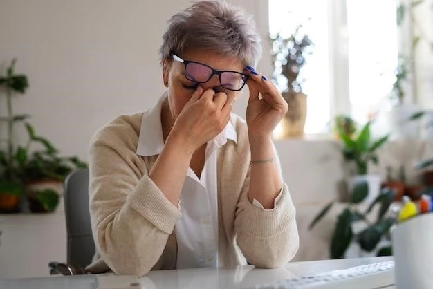
Glaucoma Iridogoniodysgenesis presents a spectrum of symptoms and effects that can significantly impact visual function and overall ocular health. Individuals with this condition may experience subtle early signs, such as intermittent blurred vision and mild discomfort, which can progress to more pronounced symptoms as the disease advances.
One of the hallmark symptoms of Glaucoma Iridogoniodysgenesis is elevated intraocular pressure (IOP), which can cause ocular pain, redness, and visual disturbances. As the optic nerve sustains damage from prolonged pressure elevation, patients may notice gradual changes in their vision, including difficulty focusing, reduced visual acuity, and an increased sensitivity to light.
Peripheral vision loss is a common consequence of untreated Glaucoma Iridogoniodysgenesis, characterized by the gradual narrowing of the visual field or the development of tunnel vision. This restriction in the field of view can impair spatial awareness, mobility, and activities of daily living, significantly impacting the quality of life of affected individuals.
Optic nerve damage resulting from Glaucoma Iridogoniodysgenesis can lead to irreversible vision loss if left unmanaged. The progressive degeneration of retinal ganglion cells, which transmit visual information from the eye to the brain, can result in permanent visual deficits and functional impairment. Timely diagnosis and intervention are crucial to mitigate these effects and preserve visual function.
Moreover, the effects of Glaucoma Iridogoniodysgenesis extend beyond vision impairment, potentially impacting psychological well-being and overall health. The chronic nature of the disease, coupled with the need for ongoing monitoring and treatment, can cause emotional distress, anxiety, and reduced quality of life for patients and their families.
By recognizing the symptoms and effects of Glaucoma Iridogoniodysgenesis, healthcare providers can intervene early, implement appropriate management strategies, and support patients in navigating the challenges associated with this rare form of glaucoma. Through comprehensive care and a multidisciplinary approach, the aim is to minimize visual morbidity, optimize patient outcomes, and enhance the overall well-being of individuals affected by this complex ocular condition.
Diagnosis
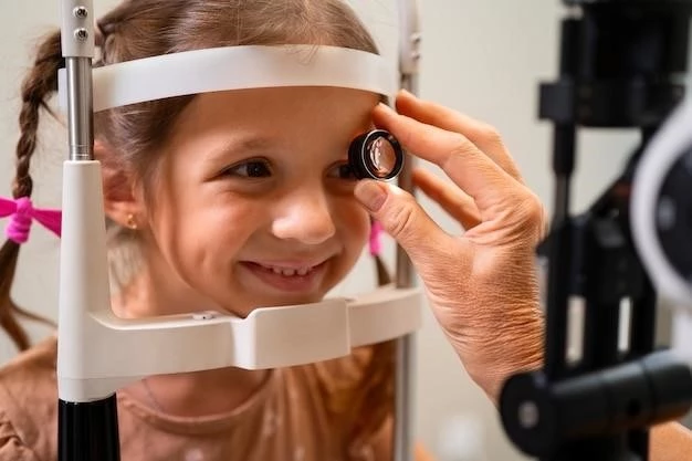
Diagnosing Glaucoma Iridogoniodysgenesis requires a comprehensive ophthalmic evaluation that includes a combination of clinical assessments, imaging studies, and intraocular pressure measurements. Healthcare providers rely on specialized techniques to detect the characteristic features of this rare form of glaucoma and differentiate it from other ocular conditions.
One of the key diagnostic procedures for Glaucoma Iridogoniodysgenesis is a thorough eye examination, during which an ophthalmologist evaluates the anterior segment of the eye to identify structural anomalies in the iridocorneal angle and surrounding tissues. Slit-lamp biomicroscopy and gonioscopy are commonly used to visualize the angle structures and assess the degree of angle closure or abnormality.
Measurement of intraocular pressure (IOP) is essential in diagnosing Glaucoma Iridogoniodysgenesis, as elevated pressure within the eye is a hallmark feature of the condition. Tonometry, a technique that quantifies the pressure inside the eye, is performed to determine the level of IOP and monitor changes over time. Persistent elevation of IOP can indicate impaired aqueous humor drainage and increased risk of optic nerve damage.
Optic nerve assessment plays a crucial role in the diagnosis of Glaucoma Iridogoniodysgenesis, as damage to the optic nerve is a significant consequence of elevated IOP. Ophthalmoscopy, optical coherence tomography (OCT), and visual field testing are employed to evaluate the structural integrity of the optic nerve head, assess retinal nerve fiber layer thickness, and detect visual field defects indicative of glaucomatous damage.
Additional imaging modalities, such as ultrasound biomicroscopy (UBM) and anterior segment optical coherence tomography (AS-OCT), may be utilized to visualize the anatomy of the iridocorneal angle and detect any structural abnormalities that contribute to impaired aqueous outflow in Glaucoma Iridogoniodysgenesis.
Given the complexity of diagnosing Glaucoma Iridogoniodysgenesis, a multidisciplinary approach involving ophthalmologists, optometrists, and imaging specialists is essential to ensure an accurate and timely diagnosis. Early detection and intervention can help prevent irreversible visual damage and enable the implementation of tailored treatment strategies to manage the condition effectively.
Treatment Options
Treating Glaucoma Iridogoniodysgenesis poses unique challenges due to its complex pathophysiology and structural abnormalities within the eye’s drainage system. Management strategies aim to reduce intraocular pressure (IOP), preserve optic nerve function, and prevent further vision loss through a combination of pharmacological, surgical, and laser interventions.
Medication therapy is often the first-line treatment for Glaucoma Iridogoniodysgenesis, with the primary goal of lowering IOP to a safe range and preventing optic nerve damage. Topical eye drops, such as prostaglandin analogs, beta-blockers, alpha agonists, and carbonic anhydrase inhibitors, are commonly prescribed to enhance aqueous outflow or reduce fluid production in the eye.
In cases where medication alone is insufficient to control IOP, surgical interventions may be recommended to improve aqueous humor drainage and lower pressure within the eye. Procedures like trabeculectomy, trabeculotomy, or goniotomy aim to create new outflow pathways or enhance existing ones to facilitate fluid drainage and reduce optic nerve strain.
Laser therapy, including selective laser trabeculoplasty (SLT) or laser peripheral iridotomy (LPI), can be utilized in the management of Glaucoma Iridogoniodysgenesis to target specific areas of the drainage angle and promote better aqueous outflow. These minimally invasive procedures help reduce IOP levels and may be beneficial for certain subtypes of the disease.
For individuals with advanced Glaucoma Iridogoniodysgenesis or refractory cases where conventional treatments are ineffective, more specialized surgical techniques like implantation of drainage devices or cyclophotocoagulation (cyclodestructive procedures) may be considered. These interventions are reserved for complex cases requiring aggressive IOP reduction.
Regular monitoring and follow-up care are essential components of managing Glaucoma Iridogoniodysgenesis to track disease progression, assess treatment efficacy, and adjust interventions as needed. Ophthalmologists play a central role in coordinating care, educating patients about their condition, and optimizing treatment plans based on individual response and visual outcomes.
By tailoring treatment approaches to the specific needs and characteristics of Glaucoma Iridogoniodysgenesis, healthcare providers aim to delay disease progression, preserve visual function, and enhance the overall quality of life for affected individuals. Collaborative decision-making and adherence to treatment regimens are key to achieving successful outcomes in the long-term management of this challenging ocular condition.
Consultation with an Ophthalmologist
Seeking consultation with an experienced Ophthalmologist is paramount for individuals diagnosed with Glaucoma Iridogoniodysgenesis. Ophthalmologists are highly trained specialists in the diagnosis, treatment, and management of ocular conditions, including rare forms of glaucoma like Glaucoma Iridogoniodysgenesis.
During the consultation, the Ophthalmologist will conduct a comprehensive evaluation to assess the specific characteristics and severity of Glaucoma Iridogoniodysgenesis in each patient. This evaluation often includes a detailed medical history review, a thorough eye examination, intraocular pressure measurements, imaging studies, and functional tests to evaluate visual function.
Based on the findings of the assessment, the Ophthalmologist will discuss treatment options tailored to the individual needs of the patient. These treatment strategies may involve a combination of medications, laser procedures, and surgical interventions to manage intraocular pressure, preserve optic nerve integrity, and prevent further vision loss associated with Glaucoma Iridogoniodysgenesis.
Furthermore, the Ophthalmologist will provide guidance on the importance of regular follow-up visits to monitor disease progression, assess treatment efficacy, and make any necessary adjustments to the management plan. Consistent monitoring is essential for optimizing visual outcomes and adjusting therapeutic interventions based on the patient’s response to treatment.
Education and counseling are integral components of the consultation process with an Ophthalmologist. Patients will receive information about their condition, including potential risks, treatment expectations, and lifestyle modifications that can support ocular health. Understanding the nature of Glaucoma Iridogoniodysgenesis empowers patients to actively participate in their care and make informed decisions about their treatment journey.
Collaboration between the patient and the Ophthalmologist is key to achieving successful outcomes in managing Glaucoma Iridogoniodysgenesis. Open communication, adherence to treatment regimens, and shared decision-making ensure that the patient receives comprehensive care, ongoing support, and access to the latest advancements in glaucoma management.
In conclusion, consulting with an Ophthalmologist specializing in glaucoma is essential for individuals with Glaucoma Iridogoniodysgenesis to receive personalized care, optimize treatment outcomes, and preserve visual function. By partnering with an experienced eye care professional, patients can navigate the challenges of this rare ocular condition with confidence and secure the best possible care for their ocular health.