Exostoses, Multiple, Type 3
Exostoses, Multiple, Type 3, also known as Hereditary Multiple Exostoses (HME3), is a genetic disorder characterized by benign bone tumors. Understanding the genetic basis and clinical features are crucial. Pediatric orthopedics considerations and distinguishing benign from malignant tumors are key topics to cover.
Introduction to Exostoses, Multiple, Type 3
Exostoses, Multiple, Type 3, also known as Hereditary Multiple Exostoses (HME3), is an autosomal dominant genetic disorder characterized by the development of multiple osteochondromas. The condition is caused by mutations in the EXT1 or EXT2 genes. Osteochondromas are cartilage-capped benign tumors that arise from the metaphyseal region of bones. While typically benign, there is a risk of malignant transformation into chondrosarcoma or osteosarcoma.
Patients with Exostoses, Multiple, Type 3 often present with multiple bony masses that can cause physical deformities, pain, and restrict joint movement. It commonly manifests in childhood and progresses as the individual grows. Timely diagnosis and management are essential to address potential complications.
Given the hereditary nature of the condition, understanding the genetic basis is crucial for proper diagnosis and risk assessment in family members. Genetic counseling plays a significant role in educating patients and their families about the inheritance pattern and the potential risk of passing the disorder to future generations.
Through advances in pediatric orthopedics, treatment options for osteochondromas have improved, focusing on alleviating symptoms, preventing complications, and monitoring for signs of malignant transformation. Differentiating between benign and malignant tumors is vital for determining the appropriate course of action and enhancing patient outcomes.
With a focus on the introduction to Exostoses, Multiple, Type 3, this article aims to provide a comprehensive overview of the disease, highlighting key aspects such as genetic underpinnings, clinical features, and pediatric orthopedics considerations. By delving into these topics, healthcare professionals, patients, and families can gain a deeper understanding of this hereditary bone dysplasia and the associated risks of sarcomas.
Understanding Schwartz-Jampel Syndrome
Schwartz-Jampel Syndrome is a rare genetic disorder characterized by skeletal abnormalities and muscle stiffness. It is distinct from Exostoses, Multiple, Type 3 but shares some similarities in terms of musculoskeletal manifestations. Patients with Schwartz-Jampel Syndrome may experience muscle weakness, joint contractures, and distinctive facial features.
The syndrome is caused by mutations in the gene responsible for producing a protein involved in muscle relaxation. This leads to muscle stiffness, particularly during childhood. Early recognition and intervention are essential to manage symptoms and improve quality of life for affected individuals.
Medical management of Schwartz-Jampel Syndrome often involves a multidisciplinary approach, including pediatric orthopedics, physical therapy, and genetic counseling. Regular monitoring for complications and adapting treatment plans based on the individual’s needs are crucial in providing holistic care.
Understanding the unique features and challenges associated with Schwartz-Jampel Syndrome is vital for healthcare professionals, caregivers, and patients alike. By raising awareness and promoting early diagnosis, the medical community can enhance support systems and interventions tailored to improve outcomes for those affected by this rare genetic disorder.
Genetic Basis of the Disease
Exostoses, Multiple, Type 3٫ is primarily caused by mutations in the EXT1 or EXT2 genes٫ which are involved in skeletal development. These genes play a crucial role in regulating the growth of bone and cartilage. Mutations in EXT1 or EXT2 lead to the formation of multiple osteochondromas٫ benign tumors composed of bone and cartilage.
The autosomal dominant inheritance pattern of the disease means that a child has a 50% chance of inheriting the mutated gene from an affected parent. Genetic testing can help identify mutations in EXT1 or EXT2, aiding in early diagnosis and risk assessment for family members. Understanding the genetic basis of Exostoses, Multiple, Type 3 is essential for personalized treatment planning and genetic counseling.
Research continues to explore the intricate mechanisms by which mutations in EXT1 and EXT2 result in the development of osteochondromas. By unraveling the genetic complexities of the disease, scientists strive to enhance diagnostic tools, therapeutic options, and genetic counseling protocols for individuals with Hereditary Multiple Exostoses.
Education about the genetic basis of Exostoses, Multiple, Type 3 empowers healthcare providers, patients, and families to make informed decisions regarding treatment and family planning. By delving into the genetic underpinnings of this hereditary bone dysplasia, the medical community can pave the way for more targeted and effective management strategies.
Clinical Features of Multiple Osteochondromas
The clinical features of Multiple Osteochondromas, a hallmark of Exostoses, Multiple, Type 3, vary but commonly include the growth of bony protrusions near the growth plates of long bones. These benign tumors may lead to skeletal deformities, restricted range of motion, and potential nerve or vascular compression.
Children with Multiple Osteochondromas may exhibit asymmetrical limb lengths, bony lumps under the skin, and joint pain. In some cases, the osteochondromas can cause pressure effects, affecting nearby tissues. Regular monitoring of the growth patterns of these benign tumors is crucial to detect any concerning changes or signs of malignant transformation.
Diagnosis of Multiple Osteochondromas often involves a combination of clinical evaluation, imaging studies such as X-rays and MRI, and genetic testing to confirm the presence of mutations in the EXT1 or EXT2 genes. Timely identification of these bony extensions is essential for early intervention and proactive management of potential complications.
Understanding the clinical features associated with Multiple Osteochondromas aids healthcare providers in devising a targeted treatment plan tailored to each patient. By recognizing the characteristic signs and symptoms of this hereditary bone dysplasia, medical professionals can ensure comprehensive care and timely interventions to optimize outcomes for individuals affected by Exostoses, Multiple, Type 3.
Specifics of MHE3 and Metaphyseal Chondrosarcoma
MHE3, a subtype of Multiple Exostoses, Type 3, is characterized by a higher risk of developing metaphyseal chondrosarcoma, a malignant tumor arising from cartilage. These chondrosarcomas are associated with EXT1 or EXT2 gene mutations and require careful monitoring due to the possibility of malignant transformation.
Metaphyseal chondrosarcomas present unique challenges in diagnosis and management, often necessitating a multidisciplinary approach involving oncologists, orthopedic surgeons, and radiologists. Treatment typically involves surgical resection of the tumor, followed by close surveillance to monitor for recurrence or metastasis.
Patients with MHE3 and metaphyseal chondrosarcoma may experience localized bone pain, swelling, and limited joint function. Imaging studies such as CT scans and PET scans are essential for accurate staging and treatment planning. Early detection and intervention play a critical role in improving outcomes for individuals with this subtype of Multiple Exostoses.
Understanding the specifics of MHE3 and metaphyseal chondrosarcoma empowers healthcare providers to tailor treatment strategies for each patient’s unique needs. By staying informed about the distinct characteristics and challenges associated with this condition, medical teams can provide comprehensive care and support to enhance the quality of life for those affected by Multiple Exostoses, Type 3.
Pediatric Orthopedics Considerations
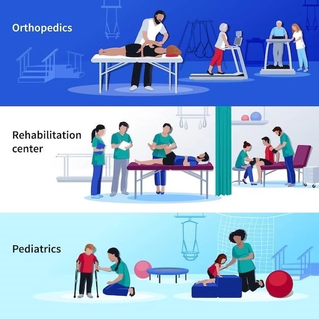
When managing Exostoses, Multiple, Type 3 in pediatric patients, several orthopedic considerations come into play. Early detection of multiple osteochondromas is crucial to monitor growth patterns, assess potential complications, and intervene proactively. Regular orthopedic evaluations are essential to track skeletal development and address any functional limitations.
Pediatric orthopedic specialists play a key role in devising treatment plans that aim to alleviate symptoms, preserve joint function, and prevent deformities associated with multiple osteochondromas. Surgical interventions may be necessary in cases where the benign tumors cause significant impairment or pose risks to adjacent structures.
Orthopedic considerations for pediatric patients with Exostoses, Multiple, Type 3 also involve the management of musculoskeletal pain, physical therapy to maintain mobility, and adaptive devices to enhance quality of life. Collaborating with physical therapists, genetic counselors, and other healthcare professionals helps ensure comprehensive care for children with this genetic disorder.
Educating parents and caregivers about the importance of regular orthopedic follow-ups, adherence to treatment plans, and recognizing warning signs of complications is paramount in optimizing outcomes for pediatric patients with Multiple Osteochondromas. By addressing orthopedic considerations early and comprehensively, healthcare providers can support the long-term musculoskeletal health of children affected by Exostoses, Multiple, Type 3.
Differentiating Benign and Malignant Tumors
Recognizing the distinction between benign and malignant tumors in the context of Exostoses, Multiple, Type 3 is crucial for appropriate management and prognosis. Benign osteochondromas, though generally harmless, can lead to discomfort and functional impairment. Monitoring their growth patterns and any changes in symptoms is essential.
In contrast, malignant transformation of osteochondromas, particularly into chondrosarcoma or osteosarcoma, presents a significant concern due to the potential for aggressive growth and metastasis. Differentiating between benign and malignant tumors involves thorough clinical evaluations, imaging studies, and histopathological analysis of tissue samples.
Features that raise suspicion for malignant transformation include rapid tumor growth, increased pain, bone destruction, and evidence of metastasis. Early detection of malignant tumors is critical for initiating timely treatment and improving outcomes. Oncologists and orthopedic surgeons collaborate to develop individualized management plans based on the tumor’s nature and extent.
Educating healthcare providers, patients, and caregivers about the red flags associated with malignant transformation helps in prompt identification and intervention. Regular surveillance imaging and follow-ups are recommended to monitor for any signs of malignancy and guide treatment decisions. By understanding the nuances of differentiating benign from malignant tumors, the medical team can ensure optimal care for individuals with Exostoses, Multiple, Type 3.
Treatment Options for Osteochondromas
When considering treatment options for Osteochondromas in the context of Exostoses, Multiple, Type 3, a multidisciplinary approach is crucial to address the complexity of these benign bone tumors. Management strategies aim to alleviate pain, improve functionality, and prevent complications associated with osteochondromas.
Observation and periodic monitoring are often recommended for asymptomatic osteochondromas that do not affect daily activities. Regular clinical assessments and imaging studies help track tumor growth and assess any potential risks of malignant transformation. In cases where benign tumors cause symptoms or skeletal deformities, surgical excision may be necessary.
Surgical intervention for symptomatic osteochondromas aims to relieve pressure on surrounding tissues, restore joint mobility, and correct deformities. Orthopedic surgeons employ advanced techniques to safely remove the bony protrusions while preserving the integrity of nearby structures. Postoperative rehabilitation and physical therapy may be included to optimize recovery and regain function.
In cases where there is concern for malignant transformation or the presence of metaphyseal chondrosarcoma, a comprehensive treatment approach involving oncologists, orthopedic surgeons, and radiologists is essential. Surgery, chemotherapy, and radiation therapy may be indicated based on the tumor’s characteristics and staging.
Educating patients and families about the available treatment options, potential risks, and expected outcomes empowers them to make informed decisions regarding their care. Collaborating with a team of specialists ensures that individuals with Osteochondromas receive personalized and comprehensive treatment tailored to their specific needs and goals.
Risks of Sarcomas in Patients with Exostoses
Patients with Exostoses, Multiple, Type 3 face inherent risks of sarcomas, particularly chondrosarcoma and osteosarcoma, arising from benign osteochondromas. Understanding these potential risks is essential for timely detection, diagnosis, and intervention to improve outcomes and prognosis.
The risk of sarcomas in individuals with Multiple Osteochondromas is relatively low but requires vigilant monitoring due to the possibility of malignant transformation. Regular imaging studies, such as MRI and CT scans, help assess the growth patterns of osteochondromas and detect any signs of malignant changes.
Early signs of sarcomas in patients with Exostoses, Multiple, Type 3 may include persistent pain٫ rapid tumor growth٫ bone destruction٫ and changes in the appearance of existing osteochondromas. Prompt evaluation by healthcare providers and oncologists is crucial to determine the nature of the tumor and initiate appropriate treatment.
Oncologists collaborate with orthopedic specialists to develop individualized treatment plans based on the type, location, and extent of the sarcoma. Surgical resection, chemotherapy, and radiation therapy may be employed to address malignant tumors and prevent further progression or metastasis.
Educating patients about the importance of regular follow-ups, self-monitoring for warning signs, and adherence to recommended surveillance protocols empowers them to take an active role in their health. By raising awareness of the risks of sarcomas in patients with Exostoses, healthcare providers can enhance detection and management strategies to safeguard the well-being of individuals affected by this genetic disorder.
Genetic Counseling for Hereditary Bone Dysplasia
Genetic counseling plays a pivotal role in managing Hereditary Bone Dysplasia such as Exostoses, Multiple, Type 3; It provides individuals and families with essential information about the genetic basis, inheritance patterns, and potential risks associated with this condition.
Individuals diagnosed with Exostoses, Multiple, Type 3 benefit from genetic counseling to understand the likelihood of passing the mutated gene to future generations. Genetic counselors guide families through the implications of autosomal dominant inheritance and help navigate decisions regarding family planning and genetic testing.

Genetic counseling sessions offer a supportive environment for patients to ask questions, express concerns, and make informed choices regarding their health and that of their family members. Understanding the genetic intricacies of Hereditary Bone Dysplasia empowers individuals to proactively manage their health and well-being.
Furthermore, genetic counselors work closely with healthcare providers to coordinate personalized care plans, facilitate genetic testing, and provide ongoing support to patients and families. By integrating genetic counseling into the management of Exostoses, Multiple, Type 3, healthcare teams can optimize care and promote informed decision-making.
Empowering individuals through genetic counseling not only aids in risk assessment and early detection but also fosters a sense of control and resilience in facing challenges associated with hereditary bone dysplasias. By embracing genetic counseling as a fundamental component of care, individuals with Exostoses, Multiple, Type 3 can navigate their genetic predisposition with knowledge and confidence.
