Introduction to Craniofacial Dysostosis
Craniofacial Dysostosis is a complex condition with variou
s causes, symptoms, and treatment approaches. Understanding
the underlying causes is crucial in managing this condition.
Definition and Overview
Craniofacial Dysostosis, also known as Crouzon syndrome, is a rare genetic disorder characterized by premature fusion of certain skull bones. It leads to abnormal facial features and may affect vision, breathing, and dental development. Early diagnosis and multidisciplinary care are vital in managing this condition.
Causes of Craniofacial Dysostosis
The causes of Craniofacial Dysostosis can be attributed to
genetic mutations affecting the development of skull bones.
Environmental factors may also contribute to the condition.
Genetic Factors
Genetic factors play a significant role in Craniofacial Dysostosis, with mutations in genes such as FGFR2 and FGFR3 being commonly associated. These mutations disrupt normal bone development٫ leading to the fusion of skull bones characteristic of this condition.
Environmental Factors
Environmental factors, such as exposure to certain toxins or medications during pregnancy, can contribute to the development of Craniofacial Dysostosis. Maternal health and lifestyle choices can also influence the risk of this condition, highlighting the complex interplay between genetics and the environment.
Symptoms and Signs of Craniofacial Dysostosis
Craniofacial Dysostosis presents with distinct facial features
such as abnormal skull shape, underdeveloped upper jaw, and
protruding eyes. Understanding these signs is crucial for early
detection and management.
Craniofacial Features
The craniofacial features of Craniofacial Dysostosis include
craniosynostosis, midface hypoplasia, and shallow eye sockets.
These characteristic abnormalities can impact both function and
aesthetics, necessitating comprehensive management approaches.
Associated Symptoms
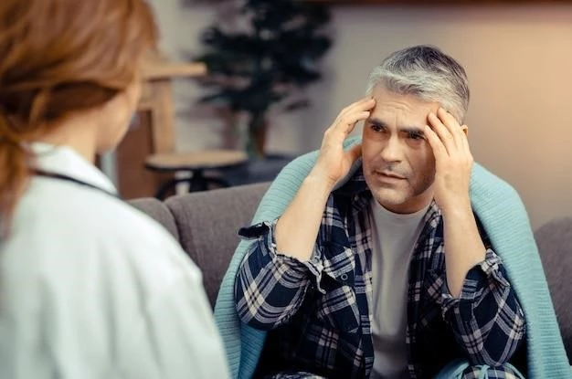
Associated symptoms of Craniofacial Dysostosis may include
breathing difficulties, sleep apnea, hearing loss, dental issues,
and potential cognitive impairments. Recognizing and addressing
these symptoms promptly is crucial for optimal patient care.
Diagnosis of Craniofacial Dysostosis
Diagnosing Craniofacial Dysostosis involves a thorough eva
luation of clinical symptoms, imaging studies, and genetic test
ing. Early and accurate diagnosis is essential for appropriate ma
nagement and treatment planning.
Physical Examination
The physical examination for Craniofacial Dysostosis involves
a detailed assessment of facial features, head shape, and skull
development. Additionally, evaluating for associated symptoms such
as respiratory issues is crucial in diagnosing this condition.
Imaging Studies
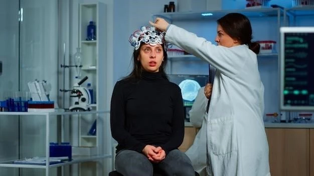
Imaging studies play a crucial role in diagnosing Craniofacial
Dysostosis by providing detailed insights into skull bone
structure and fusion patterns. Techniques such as CT scans and
MRI help in accurate assessment and treatment planning.
Treatment Options for Craniofacial Dysostosis
Treatment for Craniofacial Dysostosis includes a range of no
n-surgical and surgical interventions tailored to address specific
symptoms and improve both function and aesthetics.
Non-Surgical Approaches
Non-surgical approaches for Craniofacial Dysostosis may incl
ude orthodontic interventions, cranial molding helmets, an
d speech therapy. These techniques aim to manage symptoms an
d support normal growth and development in affected individuals.
Surgical Interventions
Surgical interventions for Craniofacial Dysostosis may involve
cranial vault remodeling, midface advancement, and orbital
reconstruction. Skilled surgical techniques aim to correct skull
deformities and enhance facial aesthetics and function effectively.
Prognosis and Complications of Craniofacial Dysostosis
The prognosis and potential complications of Craniofacial Dys
ostosis are essential considerations in the management of this
condition. Understanding these factors guides treatment and
patient care decisions.
Long-Term Outlook
The long-term outlook for individuals with Craniofacial Dysos
tosis varies depending on the severity of symptoms and the ti
mely intervention received. Regular monitoring and multidiscip
linary care are essential for optimizing outcomes over time.
Potential Complications
Potential complications of Craniofacial Dysostosis may inclu
de vision problems, hearing impairment, breathing difficulties,
dental issues, and psychological challenges. Early detection
and comprehensive management are vital in preventing and addressing these complications effectively.
Craniofacial Dysostosis in Infants and Children
Craniofacial Dysostosis in infants and children requires prompt
detection and specialized care to optimize outcomes and support
healthy growth and development. Tailored interventions are ess
ential in managing this condition at a young age.
Early Detection and Intervention
Early detection and intervention for Craniofacial Dysostosis in
infants and children are critical for ensuring appropriate manag
ement and optimal outcomes. Close monitoring, timely diagnost
ic assessments, and multidisciplinary care are essential components in addressing this condition effectively.
Impact on Growth and Development
Craniofacial Dysostosis in infants and children can affect gro
wth, feeding, speech development, and social interactions. Ear
ly and comprehensive interventions are crucial to mitigate the
impact on overall growth and development in affected individuals.
Surgical Interventions for Craniofacial Dysostosis
Surgical interventions play a crucial role in addressing the sk
ull and facial abnormalities associated with Craniofacial Dysost
osis. These procedures aim to enhance functionality and aest
hetics, improving the quality of life for affected individuals.
Types of Surgeries
Common types of surgeries for Craniofacial Dysostosis inclu
de cranial vault reconstruction, midface advancement, and jaw
realignment. These procedures are tailored to address specifi
c abnormalities and improve both function and appearance.
Post-Operative Care
Post-operative care following surgical interventions for Cranio
facial Dysostosis involves close monitoring, wound care, pain
management, and rehabilitation. Multidisciplinary support and
long-term follow-up are essential for optimal recovery and outcomes.
Research and Advances in Craniofacial Dysostosis
Ongoing research and technological advancements in Craniofacial
Dysostosis aim to improve diagnostic techniques, therapeutic
approaches, and overall outcomes for individuals affected by
this condition.
Current Research Efforts
Current research efforts in Craniofacial Dysostosis focus on
understanding genetic factors, refining surgical techniques,
exploring regenerative therapies, and improving long-term ou
tcomes. Collaborative studies aim to advance treatment strategies
and enhance quality of life for affected individuals.
Technological Innovations
Technological innovations in Craniofacial Dysostosis include adv
ances in 3D imaging, virtual surgical planning, bioengineering
solutions, and telemedicine for remote consultations. These inn
ovations enhance precision, outcomes, and access to care for affe
cted individuals globally.