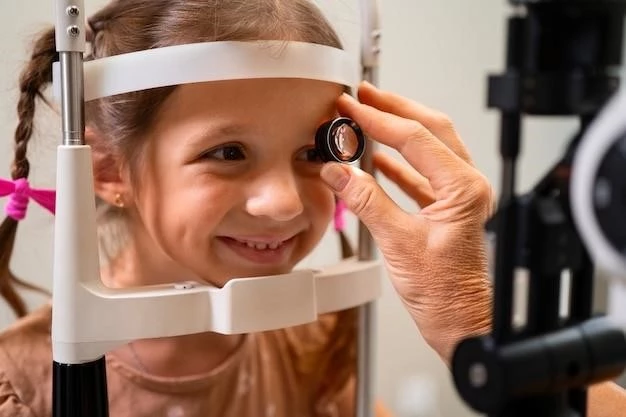
Macular Degeneration in Juveniles
When discussing juvenile-onset macular degeneration‚ it is crucial to understand the impact on central vision due to genetic mutations affecting retinal cells. This article will delve into early symptoms like blurry vision and distorted lines‚ diagnosis methods utilizing retinal imaging‚ treatment options available‚ as well as the use of vision aids and low-vision rehabilitation to manage inherited vision loss effectively.
Introduction to Juvenile-Onset Macular Degeneration
Juvenile-onset macular degeneration‚ also known as Stargardt disease‚ is a rare form of inherited vision loss that affects the retinal pigmented epithelium in the macula‚ the central part of the retina responsible for detailed‚ central vision. This condition typically manifests in children and young adults‚ causing a gradual loss of central vision over time.
Individuals with juvenile-onset macular degeneration often inherit a genetic mutation that leads to the malfunction or death of retinal cells in the macula. This mutation can impact the functioning of the retinal pigmented epithelium‚ which is essential for supporting the light-sensitive cells in the retina necessary for vision.
Unlike age-related macular degeneration‚ which affects older individuals‚ juvenile-onset macular degeneration presents unique challenges due to its early onset and potential rapid progression in younger populations. Understanding the genetic factors and mutations underlying this condition is crucial for diagnosis and targeted treatment.
Throughout this article‚ we will explore the complexities of juvenile-onset macular degeneration‚ from the genetic implications to the impact on central vision and the array of symptoms that individuals may experience. By delving into the diagnosis methods‚ treatment options‚ and strategies for managing vision loss‚ we aim to provide comprehensive insights into this condition and its management in the juvenile population.
Understanding the Retina
The retina is a crucial component of the eye responsible for converting light into neural signals that are transmitted to the brain‚ allowing us to perceive visual information. Located at the back of the eye‚ the retina consists of layers of specialized cells‚ including photoreceptor cells‚ retinal pigmented epithelium‚ and retinal ganglion cells.
Within the retina‚ the macula is a small‚ central area that plays a vital role in sharp‚ central vision required for activities like reading‚ driving‚ and recognizing faces. The macula contains a high density of cone cells‚ which are responsible for color vision and detailed visual acuity.
In juvenile-onset macular degeneration‚ the dysfunction or degeneration of the retinal pigmented epithelium‚ especially in the macula‚ can lead to a progressive loss of central vision. The impairment of this region impacts an individual’s ability to see fine details‚ resulting in difficulties with tasks that require focused vision.
Understanding the structure and function of the retina is crucial in comprehending how juvenile-onset macular degeneration affects vision. When the cells in the macula are damaged or lost due to genetic mutations‚ the clarity and sharpness of central vision diminish‚ impacting daily activities and overall quality of life.
By exploring the intricate workings of the retina‚ especially the macula‚ individuals affected by juvenile-onset macular degeneration can gain insight into the underlying mechanisms of their vision loss. This knowledge is essential for both healthcare providers involved in diagnosis and treatment and individuals navigating the challenges of living with inherited vision loss.
Genetic Factors and Mutations
Juvenile-onset macular degeneration‚ such as Stargardt disease‚ is primarily caused by genetic mutations that affect the function of retinal cells in the macula. These mutations can disrupt the normal processes within the retinal pigmented epithelium‚ leading to the degeneration and death of crucial cells responsible for central vision.
One of the most common genetic mutations associated with juvenile-onset macular degeneration is a variation in the ABCA4 gene‚ which provides instructions for producing a protein essential for normal vision. Mutations in the ABCA4 gene can result in the buildup of lipofuscin‚ a toxic byproduct that damages retinal cells‚ particularly in the macula.
Individuals who inherit these genetic mutations may experience a gradual decline in central vision due to the progressive loss of retinal cells. The inheritance pattern of juvenile-onset macular degeneration can vary‚ with some cases showing autosomal recessive inheritance‚ meaning that both parents must carry a faulty gene for their child to develop the condition.
Understanding the specific genetic factors and mutations associated with juvenile-onset macular degeneration is crucial for accurate diagnosis and targeted treatment strategies. Genetic testing can help identify the specific gene variations present in an individual‚ allowing healthcare providers to tailor interventions to address the underlying cause of vision loss.
By delving into the genetic basis of juvenile-onset macular degeneration‚ researchers aim to develop new therapies that can slow the progression of the disease and preserve remaining vision. Advancements in genetic testing and gene therapy hold promise for individuals affected by inherited vision loss‚ offering hope for improved management and potential treatments in the future.
Impact on Central Vision
Juvenile-onset macular degeneration significantly impacts central vision‚ affecting the ability to see fine details and perform tasks that require focused sight. The progressive degeneration of retinal cells in the macula leads to a decline in visual acuity‚ making it challenging to read‚ recognize faces‚ or drive.
Individuals with juvenile-onset macular degeneration often experience a gradual loss of central vision‚ with symptoms worsening over time as more retinal cells are affected by the genetic mutation. The decline in central vision can vary in severity from mild blurriness to significant visual impairment that interferes with daily activities;
One of the hallmark effects of juvenile-onset macular degeneration on central vision is the distortion of straight lines. When looking at objects or text‚ individuals may notice that lines appear wavy‚ bent‚ or distorted‚ making it difficult to discern shapes or read small print. This visual distortion is a common early symptom of macular degeneration.
The impact on central vision can have profound consequences on a person’s independence and quality of life. Tasks that rely on clear central vision‚ such as driving‚ reading‚ or recognizing faces‚ become increasingly challenging as the condition progresses. Adjusting to these changes and learning to navigate the limitations of central vision loss are important aspects of managing juvenile-onset macular degeneration.
By understanding how juvenile-onset macular degeneration affects central vision‚ individuals can proactively seek appropriate interventions and support to optimize their remaining sight. Early detection of vision changes and regular monitoring can help individuals with inherited vision loss adapt to the challenges posed by macular degeneration and make informed decisions about their eye care and overall well-being.
Early Symptoms of Juvenile-Onset Macular Degeneration

Recognizing the early symptoms of juvenile-onset macular degeneration is essential for prompt diagnosis and intervention to help preserve central vision. Individuals with this condition may initially notice subtle changes in their vision that can gradually progress over time.
One of the primary early symptoms of juvenile-onset macular degeneration is blurry vision‚ particularly in the central visual field. Objects may appear less sharp or defined‚ making it difficult to focus on details or read small print. This blurriness is often a result of the dysfunction of retinal cells in the macula.
Straight lines may also appear distorted to individuals with juvenile-onset macular degeneration. When looking at grids‚ buildings‚ or text‚ straight lines may appear wavy‚ bent‚ or irregular. This visual distortion is a common hallmark of macular degeneration and can be one of the first noticeable signs of the condition.
Difficulty adapting to changes in lighting conditions‚ such as increased sensitivity to bright lights or challenges with low-light environments‚ can also be early symptoms of juvenile-onset macular degeneration. Changes in color perception or reduced contrast sensitivity may further indicate underlying issues affecting central vision.
As juvenile-onset macular degeneration progresses‚ individuals may experience a decrease in visual acuity‚ making it harder to perform tasks that require sharp central vision. Recognizing these early symptoms and seeking evaluation by an eye care professional can aid in diagnosis and the development of a personalized management plan to address vision changes effectively.
Diagnosis and Testing
Diagnosing juvenile-onset macular degeneration involves a comprehensive eye examination and specialized testing to assess the structure and function of the retina‚ particularly the macula. Eye care professionals utilize various diagnostic tools to evaluate the extent of vision changes and identify signs of retinal degeneration.
One of the key diagnostic methods used in assessing juvenile-onset macular degeneration is retinal imaging‚ which allows healthcare providers to capture detailed images of the retina‚ including the macula. Techniques such as optical coherence tomography (OCT) provide high-resolution cross-sectional images of the retinal layers‚ highlighting any abnormalities or damage.
Fluorescein angiography is another imaging test that can help identify blood vessel abnormalities and areas of leakage in the retina‚ providing valuable information about the health of the retinal structures. This test involves injecting a fluorescent dye into the bloodstream‚ which highlights the blood flow in the retina when illuminated with a special light.
In addition to imaging tests‚ visual acuity assessments‚ color vision testing‚ and visual field tests may be conducted to evaluate the extent of vision loss and determine the impact on central and peripheral vision. Genetic testing plays a crucial role in identifying specific gene mutations associated with juvenile-onset macular degeneration.
Early and accurate diagnosis of juvenile-onset macular degeneration is essential for timely intervention and the implementation of personalized treatment plans. By combining clinical evaluations with advanced diagnostic techniques like retinal imaging‚ eye care professionals can monitor disease progression‚ tailor management strategies‚ and provide essential support to individuals affected by this rare form of inherited vision loss.
Treatment Options for Juvenile-Onset Macular Degeneration
Currently‚ there is no definitive cure for juvenile-onset macular degeneration. However‚ several treatment options are available to help manage symptoms‚ slow disease progression‚ and preserve remaining vision in individuals affected by this condition. The choice of treatment depends on the specific genetic mutation‚ stage of the disease‚ and individual circumstances.
One approach to treating juvenile-onset macular degeneration involves lifestyle modifications to support eye health‚ such as quitting smoking‚ maintaining a healthy diet rich in antioxidants and omega-3 fatty acids‚ and wearing sunglasses to protect the eyes from harmful ultraviolet (UV) rays. These lifestyle changes can help reduce oxidative stress and inflammation in the retina.
For individuals with specific genetic mutations‚ gene therapy and clinical trials may offer potential treatment options to address the underlying cause of vision loss. Gene therapy aims to introduce healthy copies of the defective gene into retinal cells‚ potentially slowing the progression of retinal degeneration and preserving vision.
In some cases‚ anti-vascular endothelial growth factor (anti-VEGF) injections may be recommended to manage macular edema or abnormal blood vessel growth in the retina. These injections can help reduce swelling‚ leakage‚ and the risk of vision loss associated with certain subtypes of juvenile-onset macular degeneration.
Low-vision aids‚ such as magnifiers‚ telescopic lenses‚ and digital devices‚ can assist individuals with juvenile-onset macular degeneration in optimizing their remaining vision and enhancing visual tasks. These devices can improve reading‚ driving‚ and performing daily activities that require precise central vision.
Collaboration with a multidisciplinary healthcare team‚ including ophthalmologists‚ optometrists‚ genetic counselors‚ and low-vision specialists‚ is essential to develop a comprehensive treatment plan tailored to the specific needs of individuals with juvenile-onset macular degeneration. By exploring a combination of lifestyle modifications‚ emerging therapies‚ and vision aids‚ individuals can better manage the challenges of living with inherited vision loss and maintain their quality of life.
Vision Aids for Managing Vision Loss
For individuals with juvenile-onset macular degeneration experiencing vision loss‚ the use of vision aids can play a crucial role in maximizing remaining eyesight and enhancing daily functioning. These aids are designed to assist individuals in performing tasks that may be challenging due to central vision impairment.
Magnifiers are commonly used vision aids that can help individuals with macular degeneration read small print‚ view objects up close‚ and perform detailed tasks. Handheld magnifiers‚ stand magnifiers‚ and electronic magnifiers with adjustable settings offer varying levels of magnification to suit different needs.
Telescopic lenses are another type of vision aid that can be beneficial for individuals with juvenile-onset macular degeneration. These lenses help with distance vision‚ allowing individuals to see objects and people clearly from afar‚ which can enhance activities like watching television‚ attending events‚ or recognizing faces across a room.
Electronic devices‚ such as tablets‚ smartphones‚ and computers‚ equipped with accessibility features like screen magnification‚ voice commands‚ and high-contrast settings‚ can aid individuals with macular degeneration in accessing digital content‚ sending messages‚ and navigating applications with ease.
Larger print books‚ audiobooks‚ voice-activated devices‚ and tactile markers can also assist individuals with juvenile-onset macular degeneration in engaging with reading materials‚ accessing information‚ and organizing their living spaces effectively. Creating a visually accessible environment is essential for promoting independence and quality of life.
Individuals with macular degeneration can benefit from working with low-vision specialists to explore the range of vision aids available and determine the most suitable options for their specific needs and preferences. By integrating vision aids into daily routines and activities‚ individuals can adapt to changes in vision‚ maintain productivity‚ and continue to participate in meaningful tasks and hobbies.
Low-Vision Rehabilitation
Low-vision rehabilitation plays a critical role in supporting individuals with juvenile-onset macular degeneration in adapting to vision loss‚ maximizing remaining sight‚ and enhancing quality of life. This comprehensive approach involves evaluating visual capabilities‚ providing training on vision aids‚ and implementing strategies to optimize functional vision.
One key component of low-vision rehabilitation is conducting a thorough assessment of an individual’s visual function‚ including visual acuity‚ contrast sensitivity‚ visual field‚ and color vision. This assessment helps low-vision specialists understand the specific challenges an individual faces and tailor interventions accordingly.
Training in the use of vision aids and assistive technologies is essential in low-vision rehabilitation. Individuals with juvenile-onset macular degeneration learn how to effectively utilize magnifiers‚ telescopes‚ digital devices‚ and adaptive tools to read‚ write‚ perform tasks‚ and engage in hobbies independently.
Environmental modifications and lighting adjustments are integral aspects of low-vision rehabilitation. Creating a well-lit environment with glare reduction‚ high-contrast colors‚ and organized spaces can enhance visibility and reduce visual strain for individuals with macular degeneration‚ making daily activities more manageable.
Another important component of low-vision rehabilitation is providing education and counseling to individuals and their families. Coping strategies‚ information on community resources‚ and emotional support play a vital role in helping individuals with juvenile-onset macular degeneration adjust to changes in vision and navigate the challenges of living with inherited vision loss.
Orientation and mobility training may be included in low-vision rehabilitation to help individuals with macular degeneration travel safely‚ navigate their surroundings‚ and maintain independence outside the home. Learning techniques for using public transportation‚ identifying landmarks‚ and negotiating obstacles can enhance confidence and mobility.
By combining personalized interventions‚ adaptive strategies‚ and ongoing support‚ low-vision rehabilitation empowers individuals with juvenile-onset macular degeneration to lead fulfilling and independent lives despite vision loss. This holistic approach addresses the physical‚ emotional‚ and practical aspects of managing inherited vision loss‚ enabling individuals to engage in activities they enjoy and maintain a sense of well-being.
