Distomatosis Disease
Distomatosis, a parasitic infection affecting farm animals, is caused by a type of fluke that primarily targets the liver and digestive system․ Understanding its transmission, symptoms, diagnosis, treatment, prevention, impact on livestock production, veterinary management, intermediate hosts, and key elements in the life cycle such as eggs and cercariae is crucial․ Moreover, exploring its zoonotic potential is essential for comprehensive knowledge․
Introduction

Distomatosis is a significant parasitic disease affecting livestock, caused by a type of fluke known as liver fluke․ These parasites target the liver and digestive system of farm animals, impacting their health and productivity; Understanding the life cycle, transmission, symptoms, diagnosis, treatment, and prevention strategies of this infection is crucial for effective management․ Additionally, the zoonotic potential of distomatosis poses a risk to human health, highlighting the importance of veterinary care and preventive measures in farm settings․
Understanding the Parasitic Fluke
The parasitic fluke responsible for distomatosis is a type of flatworm that infects farm animals, predominantly targeting the liver and digestive system․ This fluke has a complex life cycle involving intermediate hosts like snails․ The adult flukes produce eggs within the host’s body, which are excreted into the environment through feces․ The eggs hatch into larvae called cercariae, which infect the intermediate host, completing the life cycle․ Understanding the morphology, behavior, and transmission patterns of this parasitic fluke is essential for effective control and prevention of distomatosis in livestock․
Transmission of Distomatosis
The transmission of distomatosis primarily occurs through the ingestion of contaminated water or vegetation containing infective stages of the parasitic fluke․ Farm animals become infected when they ingest metacercariae present on vegetation or in water sources․ The metacercariae then develop into adult flukes within the host’s body, causing damage to the liver and digestive system․ Understanding the routes of transmission and implementing measures to control the spread of the infection are crucial aspects of managing distomatosis in livestock populations․
Symptoms in Livestock
Livestock infected with distomatosis may exhibit a range of symptoms, including weight loss, decreased appetite, decreased milk production, and general weakness․ In advanced cases, animals may show signs of abdominal pain, diarrhea, and anemia․ Severe infections can lead to liver damage and even death in some cases․ Recognizing these clinical signs and conducting proper diagnostic tests is essential for identifying and treating infected animals promptly․ Early detection of symptoms can help prevent the spread of distomatosis within livestock herds and minimize the impact on animal welfare and productivity․
Diagnosis of Distomatosis
Diagnosing distomatosis in livestock typically involves a combination of clinical evaluation, laboratory tests, and imaging studies․ Veterinary professionals may perform fecal examinations to detect the presence of fluke eggs or use advanced techniques such as serological tests to identify specific antibodies against the parasite․ Imaging modalities like ultrasonography can help visualize liver damage and the presence of adult flukes in the digestive system․ A comprehensive diagnostic approach is essential for accurate identification of distomatosis in infected animals, enabling timely treatment and management of the disease․
Treatment Options for Infected Animals
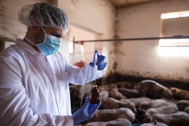
Effective treatment of distomatosis in infected animals often involves the administration of anthelmintic medications specifically targeting flukes․ These medications aim to eliminate adult parasites and control the infection․ Veterinary professionals may also recommend supportive care to manage symptoms and improve the overall health of the affected livestock․ In severe cases, additional interventions such as fluid therapy and nutritional support may be necessary to aid in the recovery process․ Regular follow-up examinations and monitoring are essential to ensure the success of treatment and prevent potential re-infections among the animal population․
Prevention Strategies
Implementing comprehensive prevention strategies is key to controlling the spread of distomatosis in livestock․ This includes maintaining good pasture management practices, ensuring access to clean water sources, and minimizing exposure to areas where intermediate hosts reside․ Regular deworming of animals with anthelmintic medications, as well as proper sanitation measures, can help reduce the risk of infection․ Quarantine protocols for introducing new animals to the herd and periodic monitoring for early detection of infections are also important preventive measures․ Educating farm staff about the disease and promoting biosecurity practices contribute to effective prevention of distomatosis․
Impact on Livestock Production
Distomatosis can have significant economic implications on livestock production due to decreased productivity and potential mortality among infected animals․ The disease may result in reduced weight gain, lower milk production, and compromised overall health, leading to financial losses for farmers․ Infected animals may require additional veterinary care, treatment, and management, further adding to production costs․ Moreover, the impact of distomatosis on herd health and reproduction can affect breeding programs and the sustainability of livestock operations․ Implementing effective control measures and early intervention strategies are essential to mitigate the negative impact of the disease on livestock production․
Veterinary Management of Distomatosis
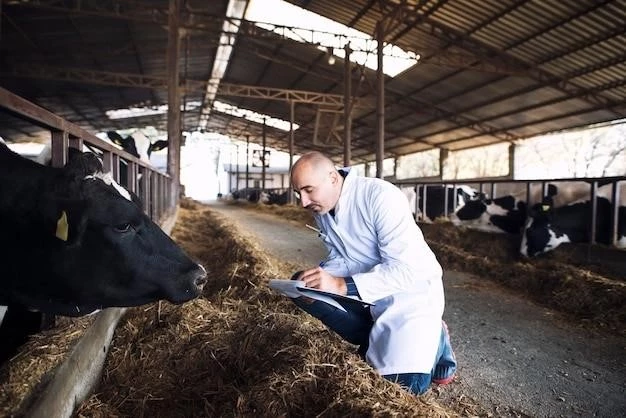
Veterinary management of distomatosis involves a multidisciplinary approach to effectively control and treat the disease in livestock populations․ Veterinarians play a crucial role in conducting regular health screenings, diagnosing infections, and prescribing appropriate treatment regimens․ They also provide guidance on preventive measures, such as deworming schedules and biosecurity protocols, to minimize the risk of transmission․ Monitoring the prevalence of distomatosis in farm animals, conducting surveillance programs, and collaborating with livestock owners are essential components of veterinary management․ By implementing comprehensive strategies, veterinary professionals aim to safeguard animal health, welfare, and productivity while ensuring the sustainability of livestock operations․
Understanding the Intermediate Hosts
In the life cycle of distomatosis, intermediate hosts play a crucial role in the transmission of the parasitic fluke․ These hosts, often snails, serve as environments for the development of the infective stages of the parasite․ Once the fluke eggs are excreted by the definitive host, they hatch into miracidia and undergo several stages of metamorphosis within the intermediate host․ The cercariae, developed from the snails, are then released into the environment, where they can infect definitive hosts such as livestock․ Understanding the biology and behavior of intermediate hosts is essential for implementing control measures to break the transmission cycle of distomatosis and reduce the prevalence of infection in farm animals․
Eggs and Cercariae⁚ Key Elements in the Life Cycle
The eggs and cercariae are crucial components in the life cycle of the distomatosis-causing fluke․ The adult flukes produce eggs within the definitive host, which are excreted into the external environment through feces․ These eggs develop into miracidia when conditions are suitable, seeking out intermediate hosts such as snails․ Within the intermediate host, the miracidia undergo multiple stages of development, eventually forming cercariae․ These cercariae are released into the surroundings where they can infect definitive hosts through ingestion․ Understanding the characteristics and behavior of eggs and cercariae is fundamental in comprehending the transmission dynamics and lifecycle of the parasitic fluke, aiding in the development of effective control strategies․
Zoonotic Potential of Distomatosis
Distomatosis, while primarily affecting livestock, also poses a zoonotic risk to human health․ In regions where the consumption of raw or undercooked animal products is common, individuals may be at risk of acquiring the infection․ Human distomatosis, caused by zoonotic fluke species, can lead to a range of symptoms including abdominal pain, diarrhea, and in severe cases, liver complications․ Proper hygiene practices, adequate cooking of food, and avoiding consumption of contaminated water are essential in reducing the zoonotic transmission of distomatosis․ Surveillance, education, and collaboration between veterinary and public health sectors are critical to mitigate the potential impact of the disease on both animal and human populations․
Conclusion
In conclusion, distomatosis is a significant parasitic disease affecting farm animals, primarily caused by liver flukes․ Understanding the transmission, symptoms, diagnosis, treatment, and prevention strategies is crucial in managing the disease effectively․ Veterinary professionals play a key role in the diagnosis and treatment of distomatosis in livestock, working to safeguard animal health and welfare․ Knowledge of the intermediate hosts, eggs, and cercariae in the life cycle of the parasite is essential for implementing control measures․ Furthermore, recognizing the zoonotic potential of distomatosis underscores the importance of interdisciplinary collaboration to address the risk to human health․ By adopting comprehensive management practices, including preventive measures and early intervention, the impact of distomatosis on both livestock production and public health can be minimized․
