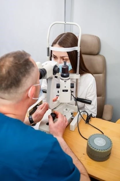
Causes of Corneal Endothelium Dystrophy
Corneal endothelium dystrophy can be caused by genetic factors, age-related degeneration,
or secondary causes such as trauma or inflammation.
Genetic Factors
Genetic factors play a significant role in corneal endothelium dystrophy, with specific gene mutations impacting the function of endothelial cells. Inherited conditions like Fuchs’ endothelial corneal dystrophy can lead to progressive damage to the cornea’s endothelium layer, affecting its ability to maintain corneal clarity.
Age-related Degeneration
Age-related degeneration can contribute to corneal endothelium dystrophy, as the endothelial cells may lose their ability to function optimally over time. Factors like oxidative stress and reduced cell turnover associated with aging can impair the endothelium’s capacity to maintain corneal transparency, leading to dystrophic changes.
Secondary Causes (e.g., trauma, inflammation)
Secondary causes like trauma or inflammation can precipitate corneal endothelium dystrophy by directly damaging the endothelial cells or disrupting the cornea’s delicate balance. Inflammatory conditions, infections, or previous eye surgeries can instigate processes leading to corneal endothelial dysfunction and dystrophic changes.
Symptoms and Diagnosis of Corneal Endothelium Dystrophy
Individuals with corneal endothelium dystrophy may experience vision disturbances and corneal edema. Diagnosis involves specialized tests like specular microscopy and pachymetry to assess the cornea’s endothelial cell layer.
Vision Disturbances
Corneal endothelium dystrophy can lead to vision disturbances such as blurred or hazy vision, glare sensitivity, and reduced visual acuity. As the condition progresses, individuals may notice worsening vision quality and difficulty with tasks like reading or driving due to corneal abnormalities affecting light transmission.
Corneal Edema
Corneal endothelium dystrophy can cause corneal edema, leading to the buildup of fluid in the cornea and resulting in swelling. This can further impair vision by distorting the cornea’s shape and transparency, contributing to symptoms like increased glare, halos around lights, and discomfort due to the altered corneal structure.
Diagnostic Tests (e.g., specular microscopy, pachymetry)
Diagnostic tests such as specular microscopy and pachymetry are utilized to assess the corneal endothelium in cases of dystrophy. Specular microscopy allows for the evaluation of endothelial cell density and morphology, while pachymetry measures corneal thickness to detect edema or abnormalities, aiding in the diagnosis and monitoring of corneal health.
Treatment Options for Corneal Endothelium Dystrophy
Treatment options for corneal endothelium dystrophy include medications, Descemet’s Stripping Endothelial Keratoplasty (DSEK),
and Endothelial Keratoplasty (DSAEK/DMEK), aimed at managing symptoms and improving corneal clarity.
Medications and Eye Drops
Medications and eye drops may be prescribed to manage symptoms of corneal endothelium dystrophy. Hypertonic saline solutions or ointments can help reduce corneal edema, while anti-inflammatory drugs can control inflammation. These treatments aim to improve corneal clarity and alleviate discomfort associated with the condition.
Descemet’s Stripping Endothelial Keratoplasty (DSEK)
Descemet’s Stripping Endothelial Keratoplasty (DSEK) is a surgical procedure used to replace damaged endothelial cells in the cornea. During DSEK, the dysfunctional endothelium is removed and replaced with a thin layer of donor tissue, promoting corneal clarity and improving vision in individuals with corneal endothelium dystrophy.
Endothelial Keratoplasty (DSAEK/DMEK)
Endothelial Keratoplasty procedures like Descemet’s Stripping Automated Endothelial Keratoplasty (DSAEK) and Descemet Membrane Endothelial Keratoplasty (DMEK) involve selective replacement of the cornea’s endothelial layer with donor tissue. These surgeries are aimed at restoring optimal corneal function and visual acuity in individuals with corneal endothelium dystrophy;
Corneal Transplant Surgery for Endothelium Dystrophy
Corneal transplant surgery, including Descemet’s Stripping Endothelial Keratoplasty, offers a comprehensive approach to treat advanced cases of corneal endothelium dystrophy.
Procedure Overview
Corneal transplant surgery for endothelium dystrophy involves removing damaged endothelial cells and replacing them with healthy donor tissue to improve corneal clarity and vision. The procedure aims to restore optimal function to the cornea while preserving as much of the patient’s corneal tissue as possible.
Recovery Process
Following corneal transplant surgery for endothelium dystrophy, patients undergo a recovery process that includes gradual improvement in vision as the new endothelial cells integrate with the cornea. Close monitoring by an ophthalmologist is essential to ensure proper healing, reduce the risk of complications, and optimize long-term visual outcomes.
Potential Risks and Complications
Corneal transplant surgery for endothelium dystrophy carries potential risks and complications, including infection, rejection of the donor tissue, increased eye pressure, and astigmatism. Close post-operative monitoring and adherence to prescribed medications are crucial to minimize these risks and optimize surgical outcomes.
Research Advances in Corneal Endothelium Dystrophy
Current research focuses on emerging therapies like tissue engineering to regenerate corneal endothelial cells for improved treatment outcomes.
Emerging Therapies (e.g., tissue engineering)
Emerging therapies such as tissue engineering hold promise in treating corneal endothelium dystrophy by developing innovative approaches to regenerate and replace damaged endothelial cells, potentially offering more targeted and effective solutions for restoring corneal clarity and visual function in affected individuals.
Clinical Trials and Studies
Ongoing clinical trials and studies are integral to advancing knowledge and treatment options for corneal endothelium dystrophy. By evaluating new therapies, surgical techniques, and treatment protocols, researchers aim to enhance outcomes and quality of life for individuals affected by this condition.
Future Directions in Treatment
Future directions in the treatment of corneal endothelium dystrophy may involve personalized medicine approaches, gene therapy, and advancements in regenerative medicine to optimize outcomes and address the underlying causes of the condition at a molecular level, paving the way for more targeted and effective treatments.

Living with Corneal Endothelium Dystrophy⁚ Tips and Support
Adopting vision rehabilitation strategies and seeking support from groups and counseling can help individuals cope with visual impairment.
Vision Rehabilitation Strategies
Implementing vision rehabilitation strategies such as low vision aids, adaptive technologies, and specialized training can enhance independence and quality of life for individuals managing corneal endothelium dystrophy. Working with low vision specialists can facilitate customized approaches to maximize visual function and everyday activities.
Support Groups and Counseling
Engaging in support groups and counseling services can provide emotional support, practical guidance, and coping strategies for individuals navigating the challenges of living with corneal endothelium dystrophy. Peer support and professional counseling can help address emotional well-being and adjustment to vision changes.
Coping Mechanisms for Visual Impairment
Developing coping mechanisms such as mindfulness techniques, maintaining a positive outlook, and adapting daily routines can help individuals effectively manage visual impairment associated with corneal endothelium dystrophy. Seeking professional guidance and incorporating assistive technologies can further enhance adaptation and quality of life.
Preventing Progression of Corneal Endothelium Dystrophy
Regular eye exams, protective eyewear, and healthy lifestyle choices are crucial in preventing the progression of corneal endothelium dystrophy.
Regular Eye Exams and Monitoring
Regular eye exams and monitoring by an eye care professional are essential to detect early signs of corneal endothelium dystrophy and track disease progression. Timely interventions based on examination findings can help preserve vision and prevent complications associated with the condition.
Protective Eyewear and Hygiene Practices
Utilizing protective eyewear, such as goggles or sunglasses, and practicing proper hygiene, including avoiding eye trauma and maintaining good eye health habits, can help prevent complications and slow the progression of corneal endothelium dystrophy.
Healthy Lifestyle Choices for Eye Health
Embracing healthy lifestyle choices such as a balanced diet rich in nutrients, regular exercise, adequate hydration, and refraining from smoking can support overall eye health and potentially slow the progression of corneal endothelium dystrophy by promoting optimal eye function and reducing oxidative stress on the cornea.
Understanding the Risk Factors of Corneal Endothelium Dystrophy
Discover the key risk factors, including age, genetics, eye trauma, surgeries, and systemic diseases.
Age and Genetics
Age and genetics are significant risk factors for corneal endothelium dystrophy, with advancing age and inherited genetic mutations contributing to the development and progression of the condition. Understanding these factors is crucial in diagnosing and managing the disease effectively.
Previous Eye Trauma or Surgeries
Previous eye trauma or surgeries can increase the risk of corneal endothelium dystrophy by damaging the delicate endothelial layer or disrupting the cornea’s structure. Understanding the impact of these factors is essential in evaluating the development and potential progression of the condition.
Systemic Diseases (e.g., diabetes, hypertension)
Systemic diseases like diabetes and hypertension are significant risk factors for corneal endothelium dystrophy, as they can impact corneal health and exacerbate endothelial dysfunction. Managing these conditions effectively is vital in minimizing the risk of developing or worsening corneal endothelium dystrophy.
