Disease ⸺ Histiocytosis X
Histiocytosis X, also known as Langerhans cell histiocytosis (LCH), is a rare disease characterized by the abnormal proliferation of immune cells called histiocytes. This condition can affect various organs such as the skin, bone, and other tissues. Understanding the different aspects of this syndrome is crucial for accurate diagnosis and effective treatment.
Introduction to Histiocytosis
Histiocytosis X, or Langerhans cell histiocytosis (LCH), is a rare disorder characterized by the overproduction and accumulation of a type of white blood cell called histiocytes. These histiocytes, part of the body’s immune system, can become abnormal and lead to inflammation and tissue damage in various organs.
In the past, histiocytosis X was classified into three distinct diseases⁚ eosinophilic granuloma, Hand-Schuller-Christian disease, and Letterer-Siwe disease. However, these terms are no longer commonly used, and the condition is now referred to as Langerhans cell histiocytosis. The exact cause of histiocytosis X is not well understood, and it is considered a disorder of immune system dysfunction.
Histiocytosis X primarily affects children, with the peak incidence occurring in those between 1 to 15 years old. However, it can also affect adults. The clinical presentation of LCH can vary widely, ranging from single organ involvement to multisystem disease. Common sites of involvement include the skin, bones, pituitary gland, liver, spleen, and lungs.
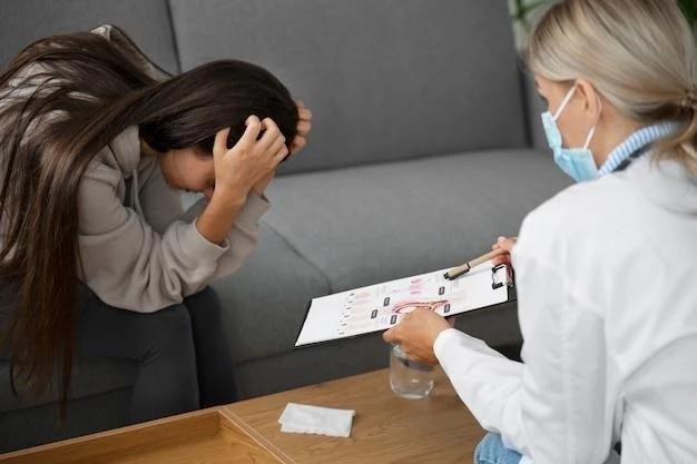
Due to the rarity and variability of histiocytosis X, diagnosis can be challenging and requires a multidisciplinary approach. Treatment options for Langerhans cell histiocytosis may include chemotherapy, steroids, and other immune-modulating medications. Biopsy of affected tissues is often necessary to confirm the diagnosis.
As a pediatric disease, histiocytosis X poses unique challenges in terms of its impact on growth, development, and long-term prognosis in children. Understanding the specific considerations for pediatric patients is essential for providing optimal care and improving outcomes in this population. Overall, histiocytosis X is a complex disease that requires careful evaluation and management to achieve the best possible results for patients.
Types and Symptoms
Langerhans cell histiocytosis (LCH) encompasses a spectrum of disease presentation, ranging from isolated lesions to disseminated multisystem involvement. The three classic forms of LCH include eosinophilic granuloma, Hand-Schuller-Christian disease, and Letterer-Siwe disease. Eosinophilic granuloma typically manifests as solitary bone lesions, commonly affecting the skull, long bones, or vertebrae.
Hand-Schuller-Christian disease presents with a triad of symptoms, including diabetes insipidus, exophthalmos, and lytic bone lesions, predominantly in the skull. Letterer-Siwe disease, the most severe form, is characterized by widespread involvement of multiple organs, leading to systemic symptoms such as fever, hepatosplenomegaly, anemia, and skin rash.
The symptoms of LCH can vary depending on the organs involved. Skin manifestations may include rashes, papules, or nodules. Bone lesions can present as pain, swelling, or pathologic fractures. In cases of pituitary gland involvement, symptoms like polyuria, polydipsia, and growth delay may arise. Pulmonary symptoms can include cough, dyspnea, and respiratory distress.
In some instances, patients with Langerhans cell histiocytosis may be asymptomatic, with the condition incidentally discovered on imaging studies performed for unrelated reasons. Due to the heterogeneous nature of LCH, a high index of suspicion is warranted, especially in cases where symptoms are nonspecific or involve multiple organ systems.
Early recognition of symptoms and prompt diagnosis are crucial for initiating appropriate treatment and preventing potential complications associated with LCH. Understanding the different types of LCH and their corresponding symptoms is essential for healthcare providers to effectively manage this complex and rare disease.
Pathophysiology and Immune Response
Langerhans cell histiocytosis (LCH) is characterized by an abnormal accumulation of Langerhans cells, a type of dendritic cell that plays a crucial role in the immune response. In LCH, these cells proliferate uncontrollably in various tissues and organs, leading to the formation of granulomas and tissue damage.
The pathogenesis of LCH is not fully understood; however, it is believed to involve dysregulation of the immune system and genetic factors. Mutations in genes such as BRAF and MAP2K1 have been identified in some cases of LCH, suggesting a genetic predisposition to the disease. These mutations can lead to the activation of signaling pathways that promote cell growth and survival.
When Langerhans cells become activated, they release pro-inflammatory cytokines and chemokines, recruiting other immune cells to the site of inflammation. This immune response contributes to the formation of granulomas, which are clusters of immune cells that surround and attempt to contain the abnormal Langerhans cells.
The dysregulated immune response in LCH can result in tissue destruction and organ dysfunction, particularly in cases of multisystem involvement. The granulomas seen in LCH can affect the function of affected organs and lead to a range of symptoms depending on the site of involvement.
Although LCH is considered a rare disease, its impact on the immune system and inflammatory pathways highlights the importance of understanding the pathophysiology of this condition. Further research into the mechanisms underlying Langerhans cell dysfunction and immune response dysregulation is essential for developing targeted therapies and improving outcomes for individuals affected by LCH.
Organ Involvement
Langerhans cell histiocytosis (LCH) can affect multiple organs throughout the body, leading to a wide range of symptoms and complications. The most commonly involved organs in LCH include the skin, bones, pituitary gland, liver, spleen, and lungs;
Skin involvement in LCH can manifest as rashes, papules, nodules, or ulcers. These skin lesions may be the initial presentation of the disease and can vary in appearance and distribution. Bone lesions are another common feature of LCH, with affected individuals experiencing bone pain, swelling, and the risk of fractures.
The pituitary gland, a crucial component of the endocrine system, can be impacted in LCH, leading to symptoms such as diabetes insipidus, growth hormone deficiency, and other hormonal abnormalities. Liver and spleen involvement in LCH can result in hepatosplenomegaly, abdominal discomfort, and potential complications related to organ dysfunction.
Lung involvement in LCH can present as a variety of respiratory symptoms, including cough, dyspnea, and lung nodules. In severe cases, lung infiltration by Langerhans cells can lead to respiratory failure and significant morbidity. Multisystem involvement in LCH necessitates a comprehensive evaluation to assess the extent of organ involvement and guide treatment decisions.
Given the diverse range of organs that can be affected in LCH, a multidisciplinary approach involving specialists from dermatology, orthopedics, endocrinology, pulmonology, and other fields is essential for the comprehensive management of this complex disease. Understanding the pattern of organ involvement in LCH is crucial for healthcare providers to formulate an appropriate diagnostic and treatment strategy tailored to the individual needs of patients.
Diagnosis
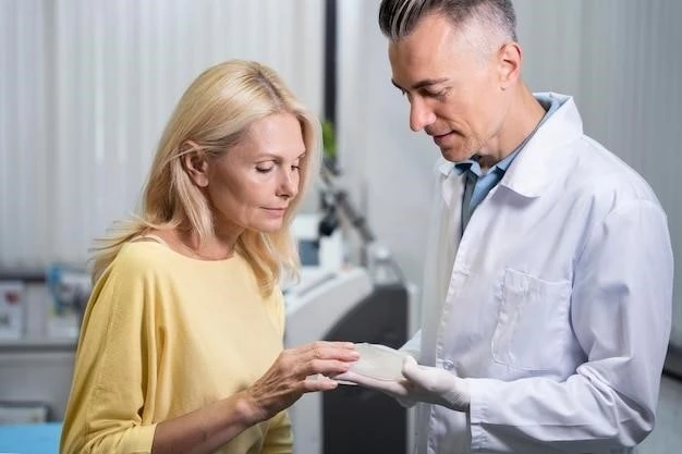
Diagnosing Langerhans cell histiocytosis (LCH) can be challenging due to its rarity and variability in presentation. A definitive diagnosis of LCH often involves a combination of clinical evaluation, imaging studies, laboratory tests, and histopathological examination of affected tissues.
Medical history plays a crucial role in the diagnostic process, as healthcare providers gather information about the patient’s symptoms, medical conditions, and family history. Physical examination may reveal characteristic skin lesions, palpable bone masses, or signs of organ involvement such as hepatosplenomegaly.
Imaging studies, including X-rays, computed tomography (CT) scans, magnetic resonance imaging (MRI), and positron emission tomography (PET) scans, are essential for detecting bone lesions, organ abnormalities, and other signs of LCH involvement. These imaging modalities help guide the selection of biopsy sites for histological confirmation of the diagnosis.
Laboratory tests, such as blood tests and urine studies, can provide additional supportive evidence for the diagnosis of LCH. Elevated levels of certain markers, such as lactate dehydrogenase (LDH) and certain cytokines, may be present in individuals with active disease. Analysis of cerebrospinal fluid may be necessary in cases of suspected central nervous system involvement.
Biopsy of affected tissues is often required to definitively diagnose LCH. Histopathological examination of biopsy samples reveals the presence of abnormal Langerhans cells, characteristic granulomas, and signs of inflammation. Immunohistochemical staining can help differentiate LCH from other histiocytic disorders and confirm the diagnosis.
Given the complexity of diagnosing LCH and the need for a multidisciplinary approach, collaboration between pediatricians, dermatologists, oncologists, pathologists, and other specialists is vital in ensuring an accurate and timely diagnosis. Early recognition and confirmation of LCH are crucial for initiating appropriate treatment and improving outcomes for individuals affected by this rare disease.
Treatment Options
Management of Langerhans cell histiocytosis (LCH) involves a tailored approach depending on the extent of disease involvement, the organs affected, and the individual’s overall health. Treatment options for LCH may include observation, chemotherapy, steroids, targeted therapies, and surgery.
Active surveillance, or watchful waiting, may be considered for asymptomatic or indolent cases of LCH, particularly when there is single-organ involvement and stable disease. Regular monitoring with imaging studies and clinical assessments is essential to detect any progression of the disease.
Chemotherapy, often with agents like vinblastine, prednisone, methotrexate, cytarabine, and cladribine, is commonly used in cases of multisystem LCH or refractory disease. Chemotherapy aims to suppress the abnormal proliferation of Langerhans cells and reduce inflammation in affected tissues.
Steroids such as prednisone may be prescribed to control inflammation and alleviate symptoms in patients with LCH. These medications can help reduce pain, improve organ function, and manage certain manifestations of the disease, particularly in cases of bone or central nervous system involvement.
Targeted therapies, including inhibitors of the MAPK pathway such as vemurafenib and dabrafenib, have shown promise in treating LCH cases with specific genetic mutations. These precision therapies target the underlying genetic abnormalities driving the abnormal proliferation of Langerhans cells.
In cases where LCH causes structural damage to bones or other tissues, surgery may be necessary to address complications such as fractures, spinal cord compression, or organ dysfunction. Surgical intervention aims to restore function, alleviate pain, and improve quality of life in affected individuals.
The selection of treatment options for LCH requires careful consideration of the individual’s age, disease severity, organ involvement, and response to previous therapies. A personalized treatment plan, often developed in collaboration with a multidisciplinary team of healthcare professionals, aims to optimize outcomes and minimize potential side effects for patients with Langerhans cell histiocytosis.
Pediatric Considerations
Langerhans cell histiocytosis (LCH) presents unique considerations when diagnosed in pediatric patients, given its predilection for affecting children and adolescents. The clinical manifestations, treatment approaches, and long-term outcomes of LCH in the pediatric population require specialized attention and expertise.
Children with LCH may present with a diverse array of symptoms depending on the organs involved and the extent of disease. Healthcare providers caring for pediatric patients must be attuned to recognizing early signs of LCH, which can sometimes mimic common childhood ailments or manifest atypically.
The impact of LCH on growth, development, and quality of life in children underscores the importance of comprehensive management strategies tailored to pediatric needs. Treatment decisions in pediatric LCH patients must carefully balance the potential benefits of therapy with the risks of long-term side effects.
Pediatric patients often require age-appropriate communication and support to understand their condition, participate in their care, and cope with the emotional aspects of having a chronic illness. Family involvement and caregiver education play a crucial role in ensuring adherence to treatment plans and optimizing outcomes.
Healthcare teams managing pediatric LCH cases should include specialists in pediatric oncology, hematology, dermatology, endocrinology, and other relevant disciplines to provide comprehensive care. Regular monitoring of growth parameters, organ function, and treatment response is essential in pediatric LCH management.
Given the potential challenges of diagnosing and treating LCH in children, ongoing research efforts focus on improving diagnostic techniques, refining treatment protocols, and enhancing supportive care measures for pediatric patients. Collaborative efforts among healthcare providers, researchers, and patient advocacy groups are vital in advancing the understanding and management of Langerhans cell histiocytosis in the pediatric population.
In conclusion, Langerhans cell histiocytosis (LCH) is a rare disease characterized by the abnormal proliferation of histiocytes, leading to inflammation and tissue damage in various organs. The complexity of LCH lies in its diverse clinical presentations, multisystem involvement, and potential long-term implications.
Diagnosing LCH requires a multidisciplinary approach, incorporating clinical evaluation, imaging studies, laboratory tests, and histopathological examination. Treatment options for LCH vary based on the extent of disease and may include chemotherapy, steroids, targeted therapies, and surgery, tailored to individual patient needs.
For pediatric patients with LCH, specialized considerations are essential to address the unique challenges associated with managing this condition in children and adolescents. Pediatric LCH cases demand a comprehensive approach that accounts for growth, development, psychosocial support, and long-term outcomes.
Ongoing research into the pathophysiology of LCH, advances in diagnostic techniques, and the development of novel treatment modalities hold promise for improving outcomes and quality of life for individuals affected by this rare disease. Collaborative efforts among healthcare professionals, researchers, and advocacy groups are critical in enhancing the understanding and management of Langerhans cell histiocytosis.
By raising awareness, promoting early detection, and individualizing treatment strategies, the medical community can work towards better outcomes and a brighter future for patients grappling with the complexities of Langerhans cell histiocytosis. Continued research and clinical advancements are imperative in advancing the field of histiocytosis and enhancing the care provided to those affected by this challenging disease.